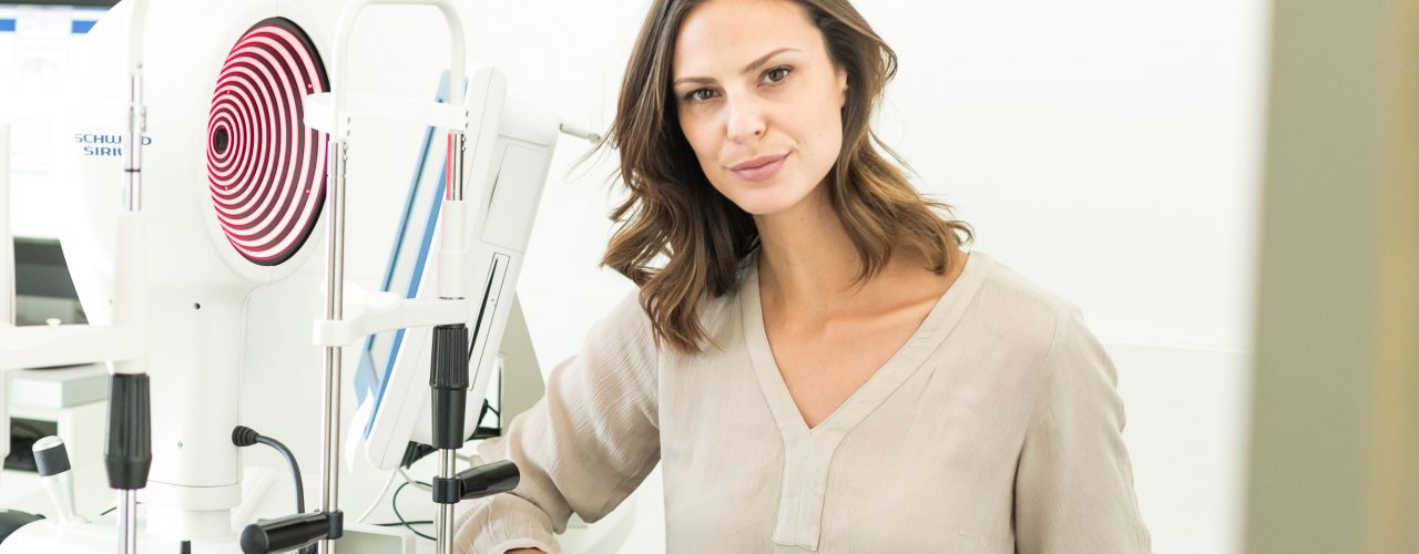
The so-called topography, which represents a digital, automated analysis of the corneal condition, is pioneering for the diagnosis of a so-called forme fruste or a progressive keratoconus.
The keratograph is an instrument for recording and evaluating the topography of the cornea. A placido disc (ring system with concentrically alternating black and white rings) is projected onto the anterior surface of the cornea, the ring-shaped reflex images are recorded with a video camera and evaluated by a computer system using a Fourier analysis.
A major advantage compared to ophthalmic measurement is the number of measurement points. In an ophthalmic measurement, only a few measuring points are recorded (two central, four peripheral). Depending on the device, 10,000 to 30,000 measuring points are recorded in the keratographs, the result being a detailed profile of the cornea. There can be selected different forms of representation. The corneal topography can be represented numerically, color-coded or as a three-dimensional flat rock.
The keratograph was originally developed for corneal surgery. In the meantime, it is also widely used as a preliminary examination as part of a cataract surgery or before a LASIK procedure, as well as in contact lens fitting.
Our cornea consists of many layers. The innermost layer is the endothelium, whose function is to keep the cornea clear. Malfunctions in this endothelial layer can lead to blurred vision. With the help of endothelial microscopy, the number of endothelial cells and the cell morphology can be assessed.
Endothelial microscopy is recommended to assess the risk of postoperative corneal irritation, such as after cataract surgery with the implantation of an intraocular lens. This examination is an optional service.
